Identification of prolonged disorders of consciousness by measuring electrical signals from neuronal networks
BEIJING, FENGTAI, CHINA, August 14, 2026 /EINPresswire.com/ — For patients with severe brain injuries, precisely identifying pDOC is a difficult task, and inaccurate identification limits how well treatments work. Current data show that even with sophisticated diagnostic instruments, false diagnoses occur in 40% of cases. This largely stems from patient factors—fatigue, sedation, or unresponsive states. Currently, physicians rely on a range of brain imaging methods as the definitive approach to detecting consciousness in patients.
The Hebei Medical University team examined multiple scientific research databases to synthesize the available evidence about pDOC management strategies. Their primary emphasis was on investigations that captured the brain’s electrical signals directly in pDOC patients, known as “local field potential.” The project, headed by Dr. Conghui Li and Dr. Yi Yang, appeared in Volume 12, article 22, on July 13, 2026, in the Chinese Neurosurgical Journal. Dr. Conghui Li stated, “Our goal is to examine how recordings of brain electrical signals aid in diagnosing pDOC and to explore the potential benefits and drawbacks of this method.”
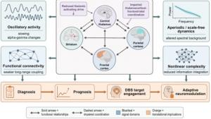
This review puts forward a model of brain communication networks to clarify how injury to the brain leads to extended unconsciousness. According to this model, brain damage destroys or harms numerous nerve cells, interrupting the transmission of electrical signals to deeper structures such as the thalamus. Consequently, interaction among key brain regions weakens, diminishing overall brain function. Supporting this view, four principal measures of brain electrical signaling activity are identified that assist researchers in gauging the patient’s level of consciousness.
In a healthy conscious brain, there is a constant, orderly, and balanced stream of activating slow signals and calming fast signals, encompassing both rhythmic and non‑rhythmic types. Notably, various parts of the active brain maintain communication with one another in a controlled, non‑chaotic manner. Another scientific theory holds that an active brain performs multiple tasks simultaneously—each region processes its own information while also keeping its communication network operational.
Severe brain injury disrupts all these balanced signaling pathways and communication networks: the fast, active brain waves transform into weaker ones, while slower signals become more predominant. Additionally, the coordinated functioning of multiple brain areas is impaired; instead of a continuous flow, the brain emits short, simple, and less organized signals. The brain’s capacity for information processing also declines during unconsciousness. Collectively, these changes reduce a person’s awareness and responsiveness.
Current studies that compare the healthy brain to the severely injured brain reveal poorer communication between the thalamus and the brain’s outer layer in individuals with pDOC. Measuring the complexity of this signaling activity—especially following brain stimulation—enables physicians to estimate the patient’s level of consciousness.
Research drawing on animal studies, anesthesia‑based investigations, and studies of brain‑injured patients has identified five additional important brain signaling activities that may improve the understanding, diagnosis, and treatment of pDOC. In severely brain‑injured patients, a lack of coordination among different brain regions and an interrupted electrical signal flow, appearing as short bursts, are observed. A failure of individual brain cells to fire in a coordinated manner in response to rhythmic signals, together with a disturbed equilibrium in brain signaling activity, is also seen in pDOC patients. Mild stimulation of the brain—particularly the thalamus—restores normal activity, so measuring the brain’s response to stimulation serves as a trustworthy method for detecting hidden awareness.
Dr. Li noted, “The research also raises important ethical issues, such as obtaining consent from family members, ensuring equity in treatment access, and clearly communicating uncertain test results to families.”
Nevertheless, more detailed investigations are needed in the future. The studies reviewed included only a small number of patients with varying types of brain injuries, and recordings were obtained under diverse conditions. Therefore, the findings cannot be reliably applied to individual patients.
In summary, directly measuring brain electrical signals from inner brain regions enables more accurate diagnoses and more tailored treatments. As Dr. Li put it, “Larger studies involving broad patient populations, hospital collaboration, and robust ethical guidelines are essential to make this diagnostic test more dependable.”
Reference
Title of original paper: Advances in local field potential research in prolonged disorders of consciousness: a narrative review
Journal: Chinese Neurosurgical Journal
DOI: https://doi.org/10.1186/s41016-026-00441-x
About the University
Hebei Medical University (abbreviated as HMU), originally called the Beiyang Medical School, ranks among the top universities in Hebei Province. Founded by Governor Li Hongzhang in Tianjin in 1894, Hebei Medical University was the first government‑established Western medical school in China. HMU offers four post‑doctoral programs—Basic Medical Sciences, Clinical Medicine, Integration of TCM and Western Medicine, and Biology—along with 37 doctoral programs and 52 master’s programs. The university runs five directly affiliated hospitals, five indirectly affiliated hospitals, five teaching hospitals, and 28 clinical practice sites. HMU serves not only as a hub for medical education and healthcare services but also as the center for medical research in Hebei Province.
Website: https://en.hebmu.edu.cn/
About Dr. Conghui Li from The First Hospital of Hebei Medical University
Dr. Conghui Li is a neurosurgeon and researcher who works in the Department of Neurosurgery at The First Hospital of Hebei Medical University, China. His research areas include disorders of consciousness (pDOC), deep brain stimulation (DBS), thalamic electrophysiology, moyamoya disease, and cerebral revascularization surgery. He has contributed to numerous peer‑reviewed publications in neurosurgery and neuroscience, covering topics such as intracranial electrophysiology, prolonged disorders of consciousness, and advanced neurosurgical procedures. His work aims to deepen understanding of brain function after severe injury and to develop improved diagnostic and therapeutic approaches for neurological conditions.
About Dr. Yi Yang from Beijing Tiantan Hospital, Capital Medical University
Professor Yi Yang, MD, serves as a neurosurgeon, researcher, and associate professor at Beijing Tiantan Hospital, Capital Medical University, where she also acts as a doctoral supervisor. Her research focuses on disorders of consciousness, deep brain stimulation, neuromodulation, brain–computer interfaces, and clinical neurophysiology. She has authored more than 60 peer‑reviewed articles and has led over 15 national and provincial research initiatives. As a Visiting Scholar at Oxford University, Dr. Yang has been recognized as a Beijing Science and Technology Rising Star and Beijing Brain Science Young Scholar. Her work merges advanced neuroscience with innovative clinical technologies to enhance the diagnosis, treatment, and rehabilitation of patients with severe neurological disorders.
Funding information
This study was funded by International (Hong Kong, Macao, and Taiwan) Science and Technology Cooperation Project (Z221100002722014), Science and Technology Innovation 2030 (2022ZD0205300), Chinese Institute for Brain Research Youth Scholar Program (2022-NKX-XM-02), National Natural Science Foundation of China (82371197), and Natural Science Foundation of Beijing Municipality (7232049).
Yi Lu
Chinese Neurosurgical Journal
email us here